
-
Table of Contents
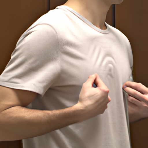
A lump on the chest wall can be a cause for concern, but not all lumps are necessarily harmful or painful. In this article, we will discuss five potential causes for a painless chest lump. It is important to note that any new or unusual lump should be evaluated by a healthcare professional to determine the underlying cause and appropriate treatment.
Lipoma: A Common Benign Tumor on the Chest Wall
A lump on the chest wall can be a cause for concern, but not all lumps are necessarily a sign of something serious. One common cause of a painless chest lump is a lipoma, which is a benign tumor that forms under the skin. Lipomas are typically soft to the touch and can move easily when pressed. They are usually painless and do not cause any other symptoms.
Lipomas are made up of fat cells and can occur anywhere on the body, including the chest wall. They are more common in middle-aged adults, but can also occur in younger individuals. The exact cause of lipomas is unknown, but they are thought to be related to genetic factors and can run in families.
While lipomas are generally harmless, they can sometimes grow larger and become more noticeable. In rare cases, they can cause discomfort if they press on nearby nerves or blood vessels. However, most lipomas do not require treatment unless they are causing significant symptoms or cosmetic concerns.
If you discover a lump on your chest wall, it is important to have it evaluated by a healthcare professional to determine the cause. They will typically perform a physical examination and may order additional tests, such as an ultrasound or biopsy, to confirm the diagnosis.
In addition to lipomas, there are several other possible causes for a painless chest lump. One possible cause is a cyst, which is a fluid-filled sac that can develop under the skin. Cysts are usually harmless and can be easily treated by draining the fluid or surgically removing the cyst.
Another possible cause is a fibroadenoma, which is a noncancerous breast tumor that can occur in both men and women. Fibroadenomas are typically firm and rubbery to the touch and can move easily. They are more common in younger women and may shrink or disappear on their own over time.
In some cases, a painless chest lump may be a sign of a more serious condition, such as a breast cancer or a metastatic tumor that has spread from another part of the body. While these are less common causes, it is important to rule them out through further testing if there are any concerning features or if the lump is growing rapidly.
In conclusion, a painless lump on the chest wall can have several possible causes, with lipomas being one of the most common. Lipomas are benign tumors made up of fat cells and are typically soft, movable, and painless. While they are generally harmless, it is important to have any new lump evaluated by a healthcare professional to determine the cause and rule out any more serious conditions. If you notice a lump on your chest wall, do not panic, but do seek medical attention for a proper diagnosis and appropriate management.
Fibroadenoma: Understanding Non-Cancerous Breast Lumps
A lump on the chest wall can be a cause for concern, but not all lumps are cancerous. One common non-cancerous lump that can occur in the breast is called a fibroadenoma. Fibroadenomas are benign tumors that develop in the breast tissue. They are usually painless and can be felt as a firm, rubbery lump.
There are several possible causes for the development of fibroadenomas. Hormonal changes during puberty and pregnancy can increase the risk of developing these lumps. Additionally, certain hormone therapies and the use of birth control pills may also contribute to their formation. It is important to note that fibroadenomas are not caused by breast cancer or any other serious medical condition.
Fibroadenomas are most commonly found in women between the ages of 15 and 35, although they can occur at any age. They are more common in women with a family history of fibroadenomas or other benign breast conditions. While fibroadenomas can occur in both breasts, they are often found in only one breast.
The diagnosis of a fibroadenoma typically involves a physical examination and imaging tests, such as a mammogram or ultrasound. In some cases, a biopsy may be performed to confirm the diagnosis. It is important to consult a healthcare professional if you discover a lump on your chest wall, as they will be able to determine the cause and provide appropriate treatment if necessary.
Treatment for fibroadenomas is not always necessary, especially if the lump is small and not causing any symptoms. In these cases, the healthcare professional may recommend regular monitoring to ensure that the lump does not change in size or shape. If the fibroadenoma is large or causing discomfort, it may be surgically removed. This procedure is typically done under local anesthesia and involves making a small incision to remove the lump.
While fibroadenomas are non-cancerous, it is still important to be aware of any changes in your breasts. Regular self-examinations and mammograms can help detect any abnormalities early on. If you notice any changes in the size, shape, or texture of a lump, or if you experience any pain or discharge from the nipple, it is important to consult a healthcare professional for further evaluation.
In conclusion, a lump on the chest wall can be a cause for concern, but not all lumps are cancerous. Fibroadenomas are a common non-cancerous breast lump that can occur in women of all ages. They are usually painless and can be felt as a firm, rubbery lump. Hormonal changes and certain medications can increase the risk of developing fibroadenomas. While treatment is not always necessary, regular monitoring and self-examinations are important to ensure that any changes are detected early on. If you discover a lump on your chest wall, it is important to consult a healthcare professional for further evaluation and appropriate treatment if necessary.
Cyst: Exploring Fluid-Filled Lumps on the Chest Wall

A lump on the chest wall can be a cause for concern, especially if it is painless. While there are various possible causes for such a lump, one common explanation is a cyst. Cysts are fluid-filled sacs that can develop in different parts of the body, including the chest wall. Understanding the causes and characteristics of cysts can help individuals better understand their condition and seek appropriate medical attention if necessary.
One possible cause of a cyst on the chest wall is a sebaceous cyst. These cysts develop when the sebaceous glands, which are responsible for producing oil to lubricate the skin and hair, become blocked. This blockage can lead to the formation of a fluid-filled sac. Sebaceous cysts are typically harmless and painless, but they can become infected if bacteria enter the cyst. In such cases, the cyst may become red, swollen, and tender.
Another potential cause of a cyst on the chest wall is an epidermoid cyst. These cysts form when cells from the top layer of the skin, known as the epidermis, multiply and accumulate within a confined space. Epidermoid cysts are usually small, round, and firm to the touch. They are typically painless unless they become infected or inflamed. In some cases, epidermoid cysts may have a central punctum, or a small opening, through which a cheesy or foul-smelling substance can be expressed.
Pilar cysts are another type of cyst that can develop on the chest wall. These cysts originate from hair follicles and are often found on the scalp, but they can also occur elsewhere on the body, including the chest. Pilar cysts are typically smooth, mobile, and round. They are usually painless unless they become inflamed or infected. Pilar cysts can sometimes be mistaken for sebaceous cysts, but they have a different origin and may require a different treatment approach.
Dermoid cysts are a less common but possible cause of a lump on the chest wall. These cysts are present at birth and develop from embryonic cells that are meant to form the skin, hair, and teeth. Dermoid cysts can contain a variety of tissues, including hair, skin, and even teeth. They are typically slow-growing and painless, but they can become infected or cause discomfort if they grow large enough to put pressure on surrounding structures.
Lastly, ganglion cysts can also occur on the chest wall. These cysts develop from the lining of a joint or tendon sheath and are filled with a thick, jelly-like fluid. Ganglion cysts are typically round or oval and can vary in size. They are usually painless unless they press on nearby nerves or cause discomfort due to their size or location.
In conclusion, a painless lump on the chest wall can be caused by various factors, one of which is a cyst. Sebaceous cysts, epidermoid cysts, pilar cysts, dermoid cysts, and ganglion cysts are all potential explanations for such a lump. While most cysts are harmless and painless, it is important to monitor them for any changes in size, shape, or symptoms. If a cyst becomes infected, inflamed, or causes discomfort, it is advisable to seek medical attention for proper evaluation and treatment.
Dermatofibroma: Causes and Characteristics of Skin Lumps on the Chest
A lump on the chest wall can be a cause for concern, especially if it is painless. While there are various reasons for the development of a chest lump, one possible cause is dermatofibroma. Dermatofibroma is a benign skin growth that commonly occurs on the chest, although it can also appear on other parts of the body.
So, what exactly is dermatofibroma? Dermatofibroma is a type of skin lesion that typically presents as a small, firm bump on the skin. It is usually brown or reddish-brown in color and can range in size from a few millimeters to a centimeter or more. While dermatofibromas are generally harmless, they can sometimes cause itching or tenderness.
The exact cause of dermatofibroma is unknown, but it is believed to be a result of an overgrowth of fibrous tissue in the skin. It is more common in women than in men and tends to occur in adults, although it can also affect children. Dermatofibromas are often found incidentally during a routine physical examination or self-examination of the skin.
There are several characteristics that can help distinguish a dermatofibroma from other types of skin lumps. One characteristic is the dimple sign, which refers to the depression that occurs when the dermatofibroma is squeezed from the sides. This is due to the fibrous tissue within the lesion. Another characteristic is the “buttonhole” or “dimpled” appearance, which is caused by the central depression of the dermatofibroma.
While dermatofibromas are generally harmless, it is important to be aware of other possible causes of chest lumps. One possible cause is a lipoma, which is a benign tumor composed of fat cells. Lipomas are usually soft and movable and can occur anywhere on the body, including the chest. Another possible cause is a cyst, which is a fluid-filled sac that can develop under the skin. Cysts are typically smooth and round and can be tender to the touch.
In some cases, a chest lump may be a sign of a more serious condition, such as a breast lump or a tumor. Breast lumps can occur in both men and women and can be a sign of breast cancer. It is important to consult a healthcare professional if you notice a lump in your chest to rule out any serious underlying conditions.
In conclusion, a painless lump on the chest wall can be a cause for concern, and one possible cause is dermatofibroma. Dermatofibroma is a benign skin growth that commonly occurs on the chest. It is important to be aware of the characteristics of dermatofibroma and other possible causes of chest lumps. While dermatofibromas are generally harmless, it is always best to consult a healthcare professional for an accurate diagnosis and appropriate treatment.
Lymphadenopathy: Unraveling Enlarged Lymph Nodes on the Chest Wall
A lump on the chest wall can be a cause for concern, especially if it is painless. While there are various possible causes for a painless chest lump, one common explanation is lymphadenopathy, which refers to enlarged lymph nodes on the chest wall. Understanding the causes of lymphadenopathy can help unravel the mystery behind a painless chest lump.
One possible cause of lymphadenopathy is infection. When the body is fighting off an infection, the lymph nodes can become enlarged as they work to filter out harmful bacteria or viruses. Infections such as tuberculosis or cat scratch disease can lead to swollen lymph nodes on the chest wall. It is important to seek medical attention if you suspect an infection as the cause of your painless chest lump.
Another potential cause of lymphadenopathy is inflammation. Inflammatory conditions such as rheumatoid arthritis or sarcoidosis can trigger the immune system to respond, resulting in swollen lymph nodes. These conditions can affect various parts of the body, including the chest wall. If you have a known inflammatory condition and notice a painless lump on your chest, it is advisable to consult with your healthcare provider.
Cancer is also a possible cause of lymphadenopathy. Lymphoma, a type of cancer that affects the lymphatic system, can cause enlarged lymph nodes on the chest wall. Breast cancer can also spread to the lymph nodes in the chest, leading to their enlargement. While cancer is a serious concern, it is important to remember that not all chest lumps are cancerous. However, it is crucial to have any new or unusual lumps evaluated by a healthcare professional.
Autoimmune diseases can also contribute to lymphadenopathy. Conditions such as lupus or Sjogren’s syndrome can cause the immune system to mistakenly attack healthy tissues, including the lymph nodes. This can result in their enlargement and the development of a painless chest lump. If you have an autoimmune disease and notice a new lump on your chest, it is important to discuss it with your healthcare provider.
Lastly, certain medications can lead to lymphadenopathy. Some drugs, such as phenytoin or allopurinol, have been associated with the enlargement of lymph nodes. If you are taking any medications and notice a painless lump on your chest, it is essential to inform your healthcare provider so they can assess whether the medication could be the cause.
In conclusion, a painless lump on the chest wall can be attributed to various causes, with lymphadenopathy being a common explanation. Infections, inflammation, cancer, autoimmune diseases, and certain medications can all contribute to the enlargement of lymph nodes on the chest. It is important to seek medical attention if you notice a painless chest lump, as a healthcare professional can help determine the underlying cause and provide appropriate treatment if necessary. Remember, early detection and intervention can greatly improve outcomes, so do not hesitate to consult with a healthcare provider if you have any concerns about a lump on your chest.
Q&A
1. Lipoma: A benign tumor made up of fat cells that can develop under the skin.
2. Sebaceous cyst: A noncancerous cyst filled with a cheesy or oily material that can form on the chest wall.
3. Fibroadenoma: A benign breast tumor that can occur in both men and women.
4. Dermatofibroma: A harmless skin growth that can appear as a lump on the chest wall.
5. Lymphadenopathy: Enlarged lymph nodes in the chest area, often caused by infection or inflammation.In conclusion, there are several potential causes for a painless lump on the chest wall. These include lipoma, sebaceous cyst, fibroadenoma, dermatofibroma, and lymphadenopathy. It is important to consult a healthcare professional for proper diagnosis and appropriate treatment if a chest lump is noticed.
Hi, I’m Pablo Garduno. I am a biohacking enthusiast, and Head Writer of SanDiegoHealth.org. I write the majority of the content on this site, and appreciate you taking the time to read my work.